 Written by
One of the defining features of biology is the ability to heal.
Compare your body to a machine, and that distinction quickly becomes clear.
Think of an automobile, for instance. Most cars are built to last around 200,000 miles — maybe 15 years with regular maintenance. After that, corrosion sets in, sensors fail, and breakdowns become nearly inevitable. Mechanical systems don’t heal. They wait for intervention, and eventually must be replaced.
The human body, by contrast, is expected to last 80, 90, even over 100 years. For many people, the idea of body repair with Stem Cell Supplements is really about giving this built-in healing system better support, so the body’s own regenerative machinery can keep up with everyday wear and tear as we age. That is with no spare parts and trillions of cells laboring under constant stress. And yet it keeps going.
The difference? Biology knows how to repair itself.
But over the years, that repair slows. We don’t bounce back the way we used to. Not because we’ve lost the will, but because the body’s systems for renewal begin to wear down.
What Are Stem Cells?
Stem cells are the body’s regenerative scriptwriters—specialized cells with the rare ability to self-renew and generate many other cell types. Under the right conditions, they rebuild tissues like bone, blood, and muscle, forming the backbone of healing and repair. At their core, stem cells serve as the body’s master resource for growth, maintenance, and regeneration.
Why Stem Cells Decline with Age
At the heart of our regenerative capacity are stem cells: highly specialized cells with the capacity to generate and replenish other cell types. When conditions are right, they can rebuild bone, blood, muscle, and more. These are the scriptwriters for regeneration. How well these cells function over time is a major determinant of how we age, which is why sustained stem cell support across aging has become a central focus in longevity research.
Yet as we age, that script starts to degrade. While some stem cell pools remain relatively stable in number, others contract markedly, and nearly all become less efficient over time [1]. Their ability to divide, differentiate, and respond to stress becomes impaired. Scientists refer to this phenomenon as stem cell exhaustion, and it’s now recognized as one of the hallmarks of aging [2]. Many people first encounter the idea of repairing the body through stem cell therapy, but there’s also growing interest in non-invasive strategies that support similar regenerative pathways from within.*
At the same time, the ones that remain may lose their sensitivity to the body’s internal cues. They circulate less effectively, misinterpret signals, or fail to act altogether. Herein lies the problem: Stem cells don’t operate in isolation. They depend on guidance. And when that guidance fades or becomes distorted, so does the body’s ability to renew itself.
Which raises a key question: If regeneration depends on instruction, can we help the body write a new script for healing?
To answer that, we’ll look at three essential phases in the body’s natural healing response:
- Mobilization: calling stem cells into action
- Communication: helping them receive and send signals
- Preservation: sustaining their capacity over time
And we’ll explore how stem cell supplements may help reinforce these phases, by supporting the clarity of the body’s regenerative dialogue.
Step 1 – Stem Cell Activation: Calling the Repair Crew
Stem cells spend most of their lives in reserve — nestled in the bone marrow, blood vessels, and fat. But when the body is confronted with physical stressors, they’re swiftly called into action (stem cell activation).
That stress triggers a cascade of chemical distress signals: cytokines, growth factors, and other molecular messengers. These signals reverberate through the body, broadcasting one clear message: help needed here.
In response, stem cells mobilize, detaching from their niches and entering the bloodstream. This marks the first step in regeneration. But like any good dispatch, mobilization depends on coordination, timing, and the right conditions.
Aptly enough, some of the most promising natural compounds found in stem cell supplements come from organisms shaped by stress. Over time, these plants and algae have developed sophisticated biochemical strategies to survive extreme environments. And in the human body, stem cell supplements may help support stem cell activation.
From the Roof of the World to the Depths of Your Bone Marrow
High in the Himalayas, where the air thins and few plants thrive, sea buckthorn flourishes via the power of chemistry. To endure the harsh conditions, it generates a dense arsenal of polyphenols and other bioactive compounds. And when we consume them, these stress-born molecules may help fortify our own resilience.
In a double-blind, placebo-controlled clinical trial [3], a standardized sea buckthorn extract (CyanthOx™) significantly increased the number of several types of circulating progenitor and stem-like cells in healthy adults. Within 1 to 2 hours of intake, circulating levels of multiple types of regenerative cells increased by ~10-15%, including cells that play key roles in vascular maintenance and tissue repair.
While transient, this mobilization suggests a potential role for sea buckthorn in enhancing the body’s readiness to respond to damage or stress.
Preliminary research also indicates that sea buckthorn compounds may influence how these cells differentiate — that is, how they develop into specific roles [4]. This hints at a two-fold effect: helping mobilize the right cells, and potentially helping them prepare for what’s next.
Awakening Stem Cells from Within
Beneath the surface of Oregon’s Klamath Lake, an ecosystem shaped by glaciers and fire, grows a unique blue-green algae: Aphanizomenon flos-aquae (AFA). Forged by millennia of environmental extremes, this organism appears to stir dormant stem cells into motion.
In a human study, an extract of AFA was shown to significantly increase circulating CD34⁺ cells — a population of early progenitors involved in the regeneration of blood, immune, and endothelial systems [5]. The effect was swift, peaking at 25% above baseline just one hour after ingestion.
How might this work? In order to mobilize, stem cells must first let go.
Stem cells are held in the bone marrow by molecular signals that act like tethers. One of the most important is SDF-1, which binds to a receptor on stem cells called CXCR4, anchoring them in place.
AFA appears to interfere with this retention system, specifically by blocking the upregulation of CXCR4, which weakens the “stay-put” signal and makes it easier for stem cells to mobilize when needed.
But mobilization is only the beginning of the story.
Step 2: Stem Cell Signaling & Communication
Stem cells are, by definition, full of potential. They can become bone, blood, muscle, or nerve. But without clear direction, that potential can go to waste. Misguided repair can lead to scarring instead of regeneration. And sometimes, stem cells simply drift without ever reaching the site of damage.
This is where signaling and communication take center stage. Just as a construction site relies on blueprints and coordination, the body depends on molecular instructions to direct its repair crew. Growth factors, guidance proteins, and intracellular messengers are all part of a complex language of regeneration.
Two ancient plants — Astragalus membranaceus and Panax notoginseng — appear to support this internal communication. Both are rich in saponins, a class of naturally soap-like molecules that can interact with cell membranes and signaling pathways, translating botanical chemistry into biological action.
Astragalus & Ginseng: Ancient Herbs Guiding Stem Cells
In Traditional Chinese Medicine, Astragalus is known as a tonic, something that strengthens the system. But modern science suggests it may occupy a more specific role: helping stem cells interpret and respond to repair cues.
Astragalus contains astragalosides, compounds endowed with a dual nature — they can mix with both water and fat. This property allows them to interact with the outer layer of cells and influence the way signaling proteins work. In essence, astragalosides help stem cells listen better and respond more accurately to the body’s repair signals.
One of these compounds, astragaloside IV, has been shown in preclinical studies to modulate a key signaling system known as Notch. Notch is basically a cellular project manager — helping stem cells decide when to divide, what type of cell to become, and where to focus their efforts. In lab studies, activating Notch supports neural stem cell differentiation — guiding more cells to become neurons [6]. In effect, it helps immature cells find their path forward.
Astragalus may also help lay the physical groundwork for regeneration.
In animal models, astragaloside IV has also been shown to support the expression of VEGF (vascular endothelial growth factor), a natural signal that tells the body to build new blood vessels [7]. Blood vessels are the highways of healing: they bring in oxygen and nutrients, carry away waste, and help stem cells reach the areas that need repair.
In this respect, Astragalus acts like both a foreman and a supply coordinator, assigning jobs to stem cells as well as making sure the tools and materials get to the site.
Exosome Messengers: Supporting Regenerative Communication
Grown in the mist-covered mountains of Southwest China, Panax notoginseng has long been treasured in Traditional Chinese Medicine as a healing root. Indeed, its very name, Panax, means “all-healing,” and its legacy as a restorative botanical spans centuries. Today, modern science is uncovering the molecular underpinnings of that reputation.
Panax notoginseng contains a distinct group of saponins called notoginsenosides, which can activate the Wnt signaling pathway — a key regulator of stem cell fate. Wnt is like a regenerative switchboard: it helps stem cells decide when to wake up, how many times to divide, and what type of tissue to become.
In a recent study, notoginsenosides significantly enhanced the activity of endothelial progenitor cells, a type of stem-like cell involved in rebuilding blood vessels [8]. These cells showed improved movement, growth, and organization into new vascular structures. Notably, when Wnt signaling was blocked, the effect was abolished, reinforcing that the pathway was essential to the response.
But Panax goes a step further: it can also help cells share regenerative programs with one another.
Healing, after all, is never a solo act. Our cells rely on constant communication to orchestrate complex repair. They coordinate via exosomes: microscopic, bubble-like packets that carry proteins, RNA, and other molecular messages. These are the body’s internal memos: telling cells when to repair, what to become, and how to support the broader healing effort [9].
Emerging research suggests that Panax notoginseng may influence the molecular content of these exosomes, helping shape what messages are sent during regeneration [10]. By modulating these tiny messages, notoginsenosides could help the body align stem cells and damaged tissues around a common goal.
Step 3: Stem Cell Preservation & Maintenance
Once stem cells are activated and guided into action, one crucial task remains: protecting their capacity to repair themselves over the long haul. Like an overworked crew, stem cells pushed too hard for too long risk burnout — or even worse, depletion.
With age, these vital cells not only decline in number, they begin to lose their sense of identity: they divide less readily, respond less precisely, and eventually struggle to maintain themselves.
Some stem cell supplements include compounds that may help preserve the regenerative script itself, allowing stem cells to stay ready even as the years pile up.
One of the most intriguing such substances emerged not from a lab…but from a bee hive.
Royal Jelly: The Queen‑Making Compound
In every honeybee hive, all larvae are born genetically identical. Any of them could become a queen. But only one will.
The determining factor is not DNA. It’s nutrition.
Or more specifically, royal jelly.
This creamy, enzyme-rich secretion is produced by nurse bees and fed exclusively to a single chosen larva. That larva doesn’t just grow. It transforms.
The resulting queen bee is larger, heavier, and biologically distinct. She lives up to 40 times longer than worker bees. While workers are sterile, she lays thousands of eggs per day. Her entire physiology is rewired, from brain chemistry to reproductive anatomy.
And none of this is driven by changes to the genetic code. Rather, it is triggered by epigenetic reprogramming — modifications in how genes are expressed, without changing the DNA itself. Royal jelly shifts methylation patterns, modifies histones, and activates developmental and longevity pathways.
And though it doesn’t bestow a royal phenotype on mammals like us, the molecular effects of royal jelly reach into deeply conserved pathways, including those that govern stem cell function, aging, and regeneration.
In one compelling study, old mice fed royal jelly maintained Pax7, a genetic marker of muscle stem cells, at levels comparable to young mice [11]. These cells are essential for muscle regeneration, and their decline is a hallmark of age-related frailty. Furthermore, the royal jelly-treated mice performed significantly better in tests of strength and coordination, despite no increase in muscle mass — suggesting that function, not just form, had been maintained through the aging process.
While the full picture is still emerging, research suggests that royal jelly can help stem cells resist aging through multiple pathways.
In lab studies, it delayed cellular senescence, the state in which cells lose their ability to divide [12], as well as helped preserve mitochondrial activity [13], the energy engine that stem cells rely upon. Royal jelly has also been shown to activate key regulators of cellular maintenance and longevity, including SIRT1 and FOXO1 [14]. These are molecules that help cells adapt to stress, repair damage, and stay biologically “young.”
In some tissues, royal jelly has even been shown to help preserve telomeres, the protective caps at the ends of chromosomes that shorten with age [15]. When telomeres erode, cells lose their capacity to replicate. Slowing down telomere shortening may be one way to protect the body’s regenerative code.
The transformation of the queen bee is far more than a biological curiosity. It is a reminder that we aren’t always bound by our starting blueprint.
In humans, of course, the effects are far subtler. Yet the principle holds: the way we age and heal is not written in stone. Nutrition, environment, and molecular signals can all shape how stem cells behave, how tissues repair, and how the body adapts to challenges.
Stem Cell Supplements & The Future of Repair
Aging may be inevitable. But the way we age is not.
Stem cells are central to that story. They are the body’s regenerative scriptwriters, carrying the capacity to rebuild what time and stress have worn down. But those instructions don’t unfold automatically. They depend on signals: when to mobilize, where to go, what to become, and how long to stay ready.
With age, those signals can become muffled or misread. But as we’ve seen, there may be ways to help clarify them.
From sea buckthorn and AFA, we see the chemistry of resilience: molecules shaped by stress that may help awaken stem cells from dormancy.
From Astragalus and Panax, we inherit ancient botanical intelligence: compounds uniquely formed to interface with cell membranes and support clearer cellular communication.
And from royal jelly, we glimpse something more profound: the possibility of epigenetic reprogramming — a way of reshaping the body’s instructions for renewal without rewriting the DNA code itself.
Together, compounds like those found in stem cell supplements could do more than preserve the regenerative script. They may offer us a chance to write a different ending.
About Qualia Stem Cell™
At this point, many people begin asking what are Stem Cell Supplements and how they differ from clinical stem cell therapies. Rather than supplying new stem cells, these formulations aim to support mobilization, communication, and preservation of the body’s existing regenerative cells.
Qualia Stem Cell™ is taken just 4 days a month, awakening the body’s natural ability to heal, recover, and renew. Its unequaled stem cell support blends 15 premium ingredients into one pioneering formula.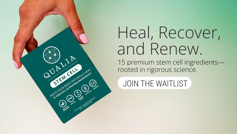
References
[1] T.A. Rando, A. Brunet, M.A. Goodell. Cell Stem Cell 2025 32(7):1038-1054.
[2] C. López-Otín, M.A. Blasco, L. Partridge, M. Serrano, G. Kroemer. Cell 2023 186(2):243-278.
[3] C. Drapeau, K.F. Benson, G.S. Jensen. Clin Interv Aging. 2019 Feb 4;14:253–263.
[4] K.H. Park, J.H. Hong, S.H. Kim, J.C. Kim, K.H. Kim, K.M. Park, Nutrients 14 (2022) 3604.
[5] G.S. Jensen, A.N. Hart, L.A.M. Zaske, C. Drapeau, N. Gupta, D.J. Schaeffer, J.A. Cruickshank, Cardiovasc. Revasc. Med. 8 (2007) 189–202.
[6] H. Hu, R. Yang, G. Jin, X. Zhang, H. Xia, Y. Xu, Evid. Based Complement. Alternat. Med. 2016 (2016) 3106980.
[7] H. Li, N. Liu, L. Yang, B. Mao, S. Ye, Int. J. Clin. Exp. Med. 9 (2016) 5709–5718.
[8] P. Zhu, W. Jiang, S. He, T. Zhang, F. Liao, D. Liu, X. An, X. Huang, N. Zhou, BMC Complement. Med. Ther. 21 (2021) 53.
[9] D. Li, D. Li, Z. Wang, J. Li, K.A. Shahzad, Y. Wang, F. Tan, Cell Biosci. 14 (2024) 105.
[10] J. Chen, Q. Zhou, Y. Lu, Front. Pharmacol. 13 (2022).
[11] N. Okumura, T. Toda, Y. Ozawa, K. Watanabe, T. Ikuta, T. Tatefuji, K. Hashimoto, T. Shimizu, Nutrients 10 (2018) 1191.
[12] M. Moriyama, Y. Miyake, N. Okumura, H. Moriyama, Biol. Pharm. Bull. 47 (2024) 2041–2049.
[13] G. Çiçek, F. Öz Bağcı, Histochem. Cell Biol. 161 (2024) 183–193.
[14] C.-M. Jiang, X. Liu, C.-X. Li, H.-C. Qian, D. Chen, C.-Q. Lai, L.-R. Shen, J. Zhejiang Univ. Sci. B 19 (2018) 960–972.
[15] S. Çakır, J. Med. Food 26 (2023) 580–585.