Written by
We’re taught to think of stress as the enemy.
But some forms of stress leave us better than before. Adversity can break you open — or make you stronger.
You know this from personal experience. Biology knows it too.
Muscles don’t grow in spite of strain. They grow because of it. Bones get denser under pressure. The immune system sharpens after exposure. Across nearly every domain of physiology, a little chaos can serve as a message: adapt, repair, rebuild.
This arc — disruption followed by repair — is a classic example of hormesis, wherein a brief dose of stress prompts long-term adaptation. It’s the logic behind strength training, cold plunges, and fasting. And increasingly, it’s being recognized as a force at work even deeper in the body — at the molecular level.
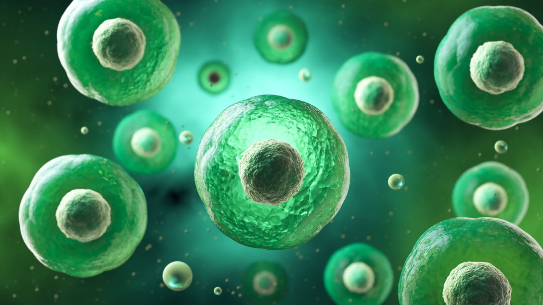
One of the most intriguing potential targets are senescent cells, aptly known as zombie cells. These are dysfunctional cells that have stopped dividing, but refuse to die. Unfortunately, senescent cells are far from passive bystanders. They’re metabolically active, secreting a cocktail of inflammatory molecules that disrupt their surroundings.
Over time, this chemical fog interferes with repair, accelerates breakdown, and impairs the body’s ability to adapt. Left unchecked, the zombie cells accumulate, creating friction in tissues that once moved easily. In skeletal muscle, their presence may be especially detrimental — interfering with recovery, performance, and muscle regeneration.
But again, stress can be a powerful signal.
Enter high-intensity interval training (HIIT). By creating controlled disruption inside the muscle itself, physical effort may help the body recognize which cells are pulling their weight… and which ones aren’t.
Let’s take a closer look at how exercise targets cellular senescence [1], and why clearing the clutter might be key to stronger, more resilient muscle.
The Burden of Cellular Dead Weight
In muscle, senescent cells, or zombie cells, do far more than linger. They interfere.
They’re like broken machinery on a factory floor: inactive, but still plugged in, leaking signals and blocking pathways. Their very presence warps the muscle’s internal landscape, disrupting the conditions that enable strength, recovery, and adaptation.
At the heart of this disruption are satellite cells. These are the muscle’s resident stem cells, responsible for repairing and rebuilding tissue after stress [2].
Satellite cells don’t work in isolation; they rely on tightly orchestrated molecular cues. But when senescent cells accumulate, those cues begin to falter. Growth signals get scrambled. Regenerative messages arrive garbled or are lost entirely. It’s like the muscle is trying to run its repair program on corrupted code.
With time, the satellite cells themselves start to lose their edge. Repeated exposure to inflammatory noise can push them toward a kind of cellular burnout — a phenomenon known as stem cell exhaustion, in which the very cells meant to renew tissue become less responsive and less capable of action.
And from there, the impact ripples outward.
Senescent cells release a steady drip of inflammatory molecules — the senescence-associated secretory phenotype, or SASP — which creates low-grade, unresolved immune activity in the tissue [3].
That background static makes it harder for muscle to recover from training, dulls the adaptive response, and slowly erodes performance capacity. Gains come more slowly. Soreness lingers longer.
Senescent cells also interfere with the metabolic machinery that powers performance [4]. They dampen insulin sensitivity, impair mitochondrial function, and slow nutrient uptake — making it harder for muscle to refuel, regenerate, and resist fatigue.
Even the extracellular matrix, the fibrous scaffolding that binds muscle fibers together and transmits force, becomes a target [5]. SASP-related enzymes can degrade or stiffen this connective tissue, fraying the muscle’s internal wiring and reducing contractile efficiency.
Individually, these effects are subtle. Together, they act like rust in a machine.
And like rust, senescent cells don’t just vanish on their own. They’re stubborn by design. But they’re not wholly immune to pressure, as we’ll soon see.
Not Just Any Workout
We have known for some time that physically active people age differently. Not just on the outside, but deep in their cells. Their immune systems stay sharper. Their blood vessels are more elastic. Even their chromosomes wear better: the protective caps on their DNA, called telomeres, erode more slowly over time.
One study, for instance, found that 50-year-old track-and-field athletes had higher telomerase activity, more telomere-stabilizing proteins, and fewer signs of senescence compared to sedentary peers [6]. On several key molecular markers of aging, the athletes more closely resembled healthy 20-year-olds than people their own age.
And a more recent study found that athletes — especially sprinters — maintain significantly higher levels of NAD⁺ and NADP⁺ in their red blood cells across the lifespan, suggesting a more youthful metabolic and redox profile [7]. NAD⁺ levels in sprinters were found to be about 32% higher than in sedentary controls.
Finally, in a landmark trial in older adults, just 12 weeks of structured exercise significantly lowered the expression of key senescence-related genes in immune cells [8]. Blood levels of senescence-associated proteins dropped as well, which was accompanied by noticeable gains in strength and stamina.
Taken together, these studies suggest that consistent training could help protect against the accumulation of senescent cells.
But here’s the catch: not all workouts create the same kind of stress. A brisk walk and an all-out sprint both count as “exercise.” But biologically, they play very different games.
Low-intensity movement relies on aerobic metabolism, the oxygen-powered engine of endurance. Push harder, though — in a sprint, a heavy lift, or a max-effort interval — and your body taps anaerobic systems. Oxygen-dependent energy falls behind. Metabolic chaos kicks in.
That chaos matters. The shift to anaerobic effort floods tissues with distinct biochemical signals, all of which may influence how the body identifies and eliminates senescent cells.
So the real question isn’t if exercise helps clear zombie cells. It’s whether intensity matters.
That’s exactly what a team of researchers in Taiwan set out to investigate [9], and their findings suggest that when it comes to cellular rejuvenation, how hard you work might matter more than how long.
Putting Intensity to the Test
To understand how exercise shapes aging at the cellular level, Taiwanese researchers recruited nine young men, and put them through a head-to-head experiment.
Rather than comparing separate groups, they used a crossover design: each participant completed both workouts, separated by at least three weeks of rest. This allowed each person to serve as their own control, filtering out individual differences in biology, fitness, and metabolism.
Before each trial, the researchers measured each participant’s maximal aerobic power, the highest output they could sustain while still relying primarily on oxygen.
Then came the workouts:
Steady-state exercise (SSE): 10 minutes at 60% of aerobic max. Think brisk cycling, challenging but controlled. Breathing increases, but the body remains composed, fueled by oxygen.
High-intensity interval training (HIIT): 15 rounds of 20-second sprints at 120% of aerobic max, with 20 seconds of rest between each. Here, the body is pushed past its oxygen ceiling. Muscles burn, breathing surges, and energy systems scramble to keep up.
Crucially, the researchers ensured that both workouts burned the same number of calories, allowing them to isolate intensity as the variable of interest.
A Drop in Cellular Age After One Workout
To determine how each workout altered the muscle at a cellular level, the researchers took biopsies from the thigh muscles at three time points: before exercise, immediately after, and again 24 hours later. These samples were then analyzed for signs of cellular aging, inflammation, and DNA damage.
The most telling results came from tracking a molecule called p16^INK4a, an established biomarker of cellular senescence. p16^INK4a functions like a cellular stop sign — its job is to prevent cells from dividing. As cells edge toward senescence, they ramp up production of this molecule, so measuring it gives us a good idea of how many senescent cells may be present.
And here’s where things got interesting: 24 hours after the high-intensity interval session, levels of the senescence marker p16^INK4a had dropped by 57%.
The steady-state session, by contrast, barely moved the needle. Despite matching the high-intensity workout for total energy output, it produced no meaningful change in senescence markers.
So what exactly triggered this drop in senescence markers?
A Controlled Burn
High-intensity interval training jolts your body into biochemical chaos. As your muscles scramble to meet energy demands, lactate builds and pH drops — a condition known as acidosis. This metabolic state is responsible for the infamous “burn” you feel during high-effort intervals.
Acidosis acts as a danger signal to the immune system, summoning macrophages into skeletal muscle [10]. In the aftermath of a hard workout, these immune cells help clear out the debris left behind by damaged muscle fibers [11]. But their role doesn’t end there — macrophages can also hunt down and eliminate senescent cells [12].
Sure enough, when the researchers compared the thigh muscle biopsies of these men, they found that immune cell infiltration into muscle ramped up 1.2-fold immediately after the high-intensity interval workout. The macrophages had arrived.
That was the first sign that the body had registered the chaos and begun to respond productively. But the immune response was just one part of the picture. Another signal was unfolding deeper inside the cells, reaching into the genome itself.
This Might Hurt a Little (But That’s the Point)
The second wave of stress came in the form of oxidative damage.
HIIT floods tissues with free radicals, disrupts cellular balance, and leads to DNA strand breaks. And indeed, when the researchers looked at muscle samples taken immediately after the HIIT session, they found a 1.3-fold increase in fragmented DNA. Steady-state exercise, by contrast, left DNA damage levels unchanged.
At first glance, this sounds like bad news. After all, DNA damage is itself a driver of cellular senescence! But the key lies in what happened next.
By the 24-hour mark, the damage was gone. DNA strand breaks had returned to baseline, suggesting that active repair was already underway. That repair process was flagged by a surge in γ-H2AX, a protein that flags broken DNA and summons the molecular tools needed to fix it.
Together, these findings reinforce a broader principle: it’s not just HIIT that can spark a senolytic response. Any workout that pushes the body into a meaningful stress-and-repair cycle may help clear senescent cells.
In fact, a separate study in young men found that a single bout of resistance training was enough to trigger a potent senolytic response [13]. Participants experienced a 48–73% drop in p16^INK4a+ endothelial progenitor cells within 48 hours. Once again, the immune system appeared to play a central role: the more macrophages infiltrated the muscle tissue, the more senescent cells disappeared.
Blocking the Burn Blocks the Benefit
High-intensity interval training is famously uncomfortable (to say the least). But what if that discomfort is the very reason HIIT works?
To find out, the same team conducted a follow-up study [14].
This time, participants completed the HIIT twice: once after taking ibuprofen, and once with a placebo. Muscle biopsies followed.
In the placebo condition — when the body was allowed to mount a normal immune response — levels of the senescence marker p16^INK4a dropped by 82% within just three hours, before gradually returning toward baseline. But when inflammation was suppressed with ibuprofen, that sharp drop was blunted.
The researchers also measured CD11b, a gene that signals the presence of activated macrophages, the immune cells that help clear out senescent cells. Because macrophages are summoned by inflammation, CD11b effectively acts as a molecular fingerprint of the inflammatory cleanup response.
After HIIT, CD11b levels fell sharply in the placebo group, signaling that the inflammatory wave had crested and the tissue was shifting into repair mode. But when participants took ibuprofen, CD11b remained static, suggesting that the body’s cleanup crew never fully mobilized — or never got the signal to stand down.
Across all samples, p16^INK4a and CD11b rose and fell in sync, suggesting that the clearance of senescent cells depends on a full and properly resolved inflammatory response.
In brief, when the inflammation was muted, the senolytic effect was too.
Growth Begins with Stress
We flinch from stress. In the gym, and in life.
But science shows us that discomfort isn’t just something to get through. It’s a catalyst for growth.
At the cellular level, high-intensity exercise sends a message. It flags what’s not working. It summons reinforcements. It clears out the dead weight. And it does all this not in spite of the strain, but because of it.
In the study we explored, that strain took the form of repeated sprints: 15 rounds of 20 seconds all-out, with just 20 seconds of rest — each interval performed at 120% of aerobic max. This kind of effort pushes the body past its oxygen limits into true anaerobic territory: time-efficient, but brutal. You don’t need lab equipment to find the zone. If you're breathless within 20 seconds, your legs are heavy, and the rest barely feels like enough to go again — you're likely where the signal starts.
Biology has a way of echoing life’s harder lessons: renewal doesn’t come through comfort, but through challenge, faced and overcome.
The Role of Senolytics
Importantly, HIIT isn’t the only challenge that delivers.
Even with the right signals, some senescent cells don’t budge. Some are deeply entrenched. They may lodge in immune-privileged tissues, or cloak themselves in molecular “don’t eat me” signals, making them tricky to clear — even for people who train hard.
That’s where senolytics come in. These compounds follow the same principle as HIIT but act through a different pathway. Two of the best-studied are fisetin and quercetin, plant compounds found in foods like strawberries, apples, and onions. Instead of taxing the whole system, they create selective pressure inside dysfunctional cells, pushing them past their tipping point. Healthy cells endure, while the zombie cells self-destruct.*
Senolytics don’t replace exercise. But they follow the same biological script — using targeted stress to spark renewal. For those cells that are too stubborn to yield, senolytics help shift the balance toward regeneration, resilience, and repair.*

* These statements have not been evaluated by the Food and Drug Administration. The products and information on this website are not intended to diagnose, treat, cure or prevent any disease. The information on this site is for educational purposes only and should not be considered medical advice.
References
[1] X. Zhang, D.A. Englund, Z. Aversa, S.K. Jachim, T.A. White, N.K. LeBrasseur, Exerc. Sport Sci. Rev. 50 (2022) 213–221.
[2] W. Chen, D. Datzkiw, M.A. Rudnicki, Open Biol. 10 (2020) 200048.
[3] S. Lopes-Paciencia, E. Saint-Germain, M.-C. Rowell, A. Fernández Ruiz, P. Kalegari, G. Ferbeyre, Cytokine 117 (2019) 15–22.
[4] A. Podraza-Farhanieh, R. Spinelli, F. Zatterale, A. Nerstedt, S. Gogg, M. Blüher, U. Smith, Mol. Metab. 95 (2025) 102130.
[5] G. Chinvattanachot, D. Rivas, G. Duque, Ageing Res. Rev. 102 (2024) 102589.
[6] C. Werner, T. Fürster, T. Widmann, J. Pöss, C. Roggia, M. Hanhoun, J. Scharhag, N. Büchner, T. Meyer, W. Kindermann, J. Haendeler, M. Böhm, U. Laufs, Circulation 120 (2009) 2438–2447.
[7] B. Pospieszna, K. Kusy, E.M. Slominska, J. Zieliński, M. Ciekot-Sołtysiak, BMC Sports Sci. Med. Rehabil. 16 (2024) 216.
[8] D.A. Englund, A.E. Sakamoto, C.M. Fritsche, A.A. Heeren, X. Zhang, B.R. Kotajarvi, D.R. Lecy, M.J. Yousefzadeh, M.J. Schafer, T.A. White, E.J. Atkinson, N.K. LeBrasseur, Aging Cell 20 (2021) e13415.
[9] W. Jean, Y. Hsieh, L. Lai, L. Dewi, Y. Liao, M. Ye, S. Yu, C. Kao, C. Huang, C. Kuo, Aging (Albany NY) 15 (2023) 765–776.
[10] G. Paulsen, R. Crameri, H.B. Benestad, J.G. Fjeld, L. Mørkrid, J. Hallén, T. Raastad, Med. Sci. Sports Exerc. 42 (2010) 75–85.
[11] J.G. Tidball, M. Wehling-Henricks, J. Physiol. 578 (2006) 327–336.
[12] M.M. Kay, Proc. Natl. Acad. Sci. U.S.A. 72 (1975) 3521–3525.
[13] C. Yang, Y. Jiao, B. Wei, Z. Yang, J.F. Wu, J. Jensen, W.H. Jean, C.Y. Huang, C.H. Kuo, Aging (Albany NY) 10 (2018) 1356–1365.
[14] W.H. Jean, Y.C. Lin, P.Y. Ang, K. Goto, C.A. Lin, L. Dewi, Y.C. Liao, C.Y. Huang, C.H. Kuo, Aging (Albany NY) 16 (2024) 8599–8610.